This product can work on multiple pathological conditions to differentiate between control and disease conditions based on an entirely non-invasive environment
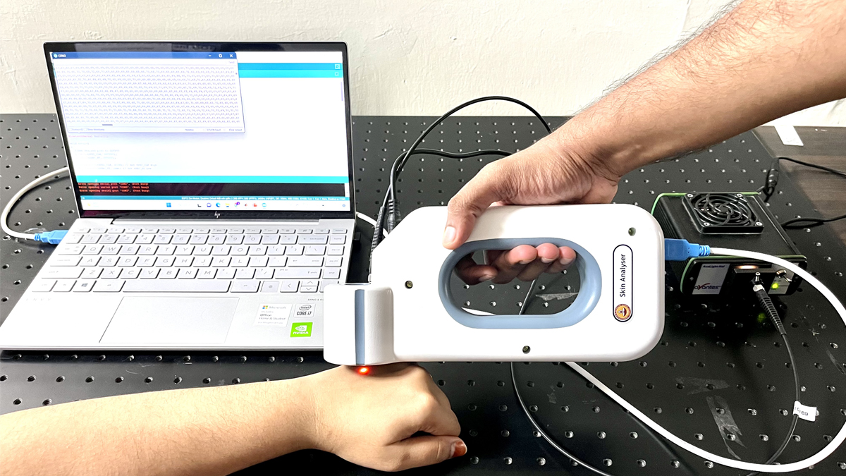
Indian Institute of Technology Madras (IIT Madras) researchers have developed a portable device that can assess alterations in skin conditions up to a depth of 2 mm. A patent was granted recently to the research team.
Diseases such as scleroderma, diabetes, and rheumatoid arthritis have altered microcirculation modulated by skin optical scattering during disease progression and can be picked up by the instrument to differentiate from the controls.
The initial set of clinical trials has been completed at SRMC, Chennai, with promising results. The final instrument has been fabricated through Design Alpha, a private firm based in Kerala.
This product is the first instrument known to date that can work on multiple pathological conditions to differentiate between control and disease conditions based on an entirely non-invasive environment.
Prof. N. Sujatha, Department of Applied Mechanics and Biomedical Engineering, IIT Madras, led the research project. This project was taken up by researchers from Biophotonics Lab, IIT Madras, in collaboration with the Department of Dermatology, SRMC Chennai, under the Biomedical Device and Technology Development initiative of the Department of Science and Technology (DST), Government of India.
Elaborating on the unique aspects of this device, Prof. N. Sujatha, Department of Applied Mechanics and Biomedical Engineering, IIT Madras, said, “Through this device, we are looking at the epidermal and dermal layers of the skin tissue embedded with microcirculatory bed. Any alterations in microcirculation caused by various disease conditions can indicate the disease progression. The developed device can look at the overall microcirculatory marker profile in the back scattered light from the skin tissue. The device would further aid the cosmetic industry in testing the effectiveness of products meant for skin rejuvenation. The device would also find use in monitoring laser-based therapeutic procedures for improving skin health.”
Regarding a timeline for the commercialization of this technology and implementation in the field, Prof. N. Sujatha added, “Initial set of trials gave promising results. These trials need to be extended to a large scale to make more generalized predictions and disease staging. Further, the device needs to be tested for various skin rejuvenation applications. A real-time version of the device is under development and expected soon.
The alterations in microcirculation are picked up using the instrument through the optical response of the tissue and further subjected to processing and classification using developed algorithms.
The approach developed by IIT Madras researchers can be applied to different pathologies after carefully studying the conditions and modifying the instrumentation and algorithms.
The Key advantages of this IIT Madras technology over existing technologies include
– Portability
– Real-time measurement
– Non-invasiveness
– Assessing multiple pathologies/skin conditions
Understanding tissue pathology and its correlation to tissue optical response is critical in developing pain-free optical biopsy techniques. In addition to increasing patient comfort, such practices offer real-time and portable solutions that are easy to use and do not require a skilled technician. Once optimized, these techniques can potentially eliminate the risk of sampling errors and excessive bleeding during sample collection compared to their tissue biopsy counterparts, the currently practiced gold standards.


