IIT Madras researchers developed this tool using deep learning and traditional machine learning.
Indian Institute of Technology Madras researchers use a combination of deep learning techniques and traditional machine learning to develop a tool that can diagnose cancer by looking at whole slide images of the tumour. The tool has been tested on datasets of breast, liver and colon cancer tissue images. It is economical in terms of the time required to process the images and analyse them.
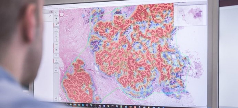
Digital histopathology
Traditionally, histopathologists slice the tumour tissue into approximately 20-micron-thick slices which they put on slides. They look at enlarged images of the slides and go over it cell by cell to manually classify it as cancerous or otherwise. This is highly time-consuming, and it is to alleviate this burden that digital histopathology developed. In this, after preparing the slides, the entire slide is scanned using a high-resolution microscope and digitised. This is then analysed using computerised tools.
This digital process has its own challenges. The first of this is the sheer size of the scanned images which can run into gigabytes.
A single image could be 100,000 by 100,000 pixels large – compare this with a shot generated by a smart phone which is about 7,000 by 7,000 pixels in size,” says Ganapathy Krishnamurthy of the Engineering Design Department of IIT Madras in an email, giving an idea of the challenge to computer time posed by this task.
The technique posed further challenges, such as insufficient training data and the variation in staining across labs.
Also extracting clinically relevant information, that is, directly inferring fine-grained disease classification using deep learning alone was turning out to be prohibitively expensive in terms of computational time.
So, the researchers used a combination of deep learning and traditional machine learning.
Hybrid approach
“We have used deep learning for classifying the tissue regions into normal and cancerous tissues from the Whole Slide Image. After this step, important patterns called features are computed using the output of the deep learning algorithm and used in a conventional machine learning algorithm for fine grained disease classification,” says Prof. Krishnamurthy. The details have been published in a paper in Scientific Reports.
To test the algorithms, the researchers enrolled in three open challenges – the CAMELYON17 challenge, the DIGESTPATH 2019 challenge and the PAIP 2019 challenge. “This paper takes data from three different challenges. In two of these, we were placed in the top 3 performing methods and in one challenge (CAMELYON) we were placed in the top 10 out of several hundred entries,” explains Balaji Srinivasan from the Mechanical Engineering Department of IIT Madras, who is an author of the paper. The technology is ready for deployment, say the authors. “We have a startup that can provide this solution and we are looking for potential healthcare partners,” says Prof. Srinivasan.
Original News Link
https://www.iitm.ac.in/happenings/press-releases-and-coverages/ai-tool-simplify-identification-cancerous-tissue-tumours


